Friday, March 25, 2011

Additional Resources for Hand, Wrist, Forearm Elbow and Shoulder Problems


Thumb Sprains


Thumb Sprains 
 
What are thumb sprains?

A sprain is an injury to a ligament. Ligaments are the connective tissues that connect bones to bones across a joint. The most common thumb sprain involves the collateral ligaments of the Metacarpophalangeal (MCP) joint.
 
How do thumb sprains occur?

These types of injuries are common in sports and falls on to an outstretched hand. The thumb is jammed into another player, the ground, or the ball. The thumb may be bent in an extreme position, causing a sprain. The thumb will usually swell and may show bruising. It is usually very painful to move.
 
What are the most common types of thumb sprains?

The most common ligament to be injured in the thumb is the ulnar collateral ligament (see Figure 1). Injury to this ligament is sometimes called “skier’s thumb” because it is a common skiing injury. It occurs when the skier falls and the pole acts as a fulcrum in the hand to bend the thumb in an extreme position. This ligament may also be injured by jamming the thumb on the ground when falling or by jamming the thumb on a ball or other player. The radial collateral ligament  may also be injured. The radial ligament is much less commonly injured than the ulnar collateral ligament 
(see Figure 1). For many years an injury to the ulnar collateral ligament of the thumb MCP joint has also been called "gamekeepers thumb" after the chronic injury to that ligament suffered by a gamekeeper  reportedly this phrase has been attributed to
 
What are some signs that this injury may have occurred?
 
Characteristic signs include pain, swelling, and bruising around the thumb , and especially over the MCP joint of the thumb. The patient will often manifest a weakened ability to grasp objects or perform such tasks as tying shoes and tearing a piece of paper. Other complaints include intense pain experienced upon catching the thumb on an object, such as when reaching into a pants pocket.
 
How are thumb sprains treated?

X-rays are usually taken to make sure the bones of the thumb and hand are not fractured. Sometimes "stress" X-rays are also used. Your doctor will then examine the thumb to determine whether the ligament is torn. If the ligament is partially torn, it is usually treated in a cast or splint. Radial collateral ligament injuries are frequently treated this way as well. The end of a completely torn ulnar collateral ligament often gets trapped behind a tendon. This "trapping" of the ligament in the edge of the aponeurosis is known as a Stener Lesion.  Complete ulnar collateral ligament tears are most commonly treated with surgery to repair the ligament. The presence or absence of a Stener Lesion is at times used to determine whether surgery is needed but typically complete lesions or ligament tears require surgery.  Sometimes the remaining ligament tissue is of poor quality and the ligament must be reconstructed with a tendon or ligament graft. 
A partial strain that does not require referral can be treated with either a thumb spica cast or a gamekeeper's thumb splint for four to six weeks. Active range of motion exercise is started at three to four weeks, with gradual return to full activity.
 
Chronic Injuries

The term “chronic” refers to an old injury of greater than one or two months duration. In this case, the joint may be unstable with symptoms of pain, especially with pinching. The joint may feel loose and strength may be decreased. These injuries may be treated by reconstruction of the ligament, or joint fusion if arthritis is present. Untreated tears can cause disabling instability of the hand, since the ulnar collateral ligament stabilizes the first MCP joint when the thumb is adducted against the index and middle fingers when gripping objects. Reconstruction typically involves taking a small piece of tendon tissue from the wrist to be placed into the thumb joint.
 
Associated Injuries

On occasion, fractures may occur along with thumb sprains. These may require additional surgery with repair using metal pins, screws, or plates. Cartilage damage may occur as well which does not show up on x-ray. This occasionally results in long-term pain and eventual arthritis. Some patients may benefit from cortisone injections or eventual surgery.
 
What happens and what should I expect after surgery?
 
A cast is placed on your thumb, wrist and forearm for approximately 4 weeks.  Your thumb tip and your fingers are left free.  After the cast is removed, the pin that holds your joint in place is typically removed in the office. You then go to hand therapy and are fitted for a removable splint that is very similar in size and shape to your cast. However you can remove this splint for exercises and range of motion.  At first you will remove the splint under the supervision of the therapist. As you progress you will be instructed by the therapist to remove the splint in controlled situations for exercise and use.  It still takes another month at least before you can use your hand without limits or splinting.  Some take longer, others go faster, but overall you have to consider it to be a 2 -3 month process.
Failure to wear a cast, and then a splint and deciding not to go to therapy can limit or compromise your result.  In general some loss of motion of the thumb occurs but the goal is to have a stable thumb joint for activity.

Bite Wounds of the HAND

Animal and Human Bites of the Hand
Bites are extremely common and can cause significant pain and other problems, especially when associated with an infection. Early recognition of warning signs and appropriate treatment are key in minimizing potential problems from the bite.
When an animal bites, bacteria from its mouth can contaminate the wound. These bacteria may grow within the wound and cause an infection. The consequences of infection range from mild discomfort to life-threatening complications.
Many factors may contribute to the infection, including the type and location of the wound, pre-existing health conditions in the bitten person that impair immunity, such as diabetes, HIV, etc., the extent of delay before treatment, the presence of a foreign body in the wound, and the animal causing the bite.
Animal Bites
There are as many as three million animal bites in the United States each year. Dogs are responsible for most animal bites in this country (up nullto 90%), with cat bites accounting for about 5% of such injuries. Other biting animals include rodents (at least 2%), rabbits, ferrets, farm animals, monkeys, and reptiles.
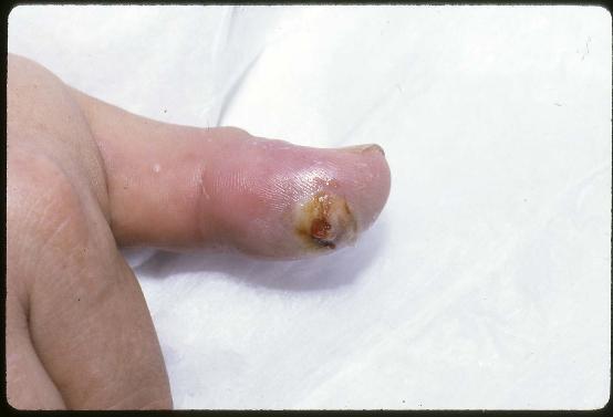
Animal bites to the hand most frequently occur on the fingers of the dominant hand of children between the ages of 5 and 14. Women are bitten more frequently by cats, and men by dogs. Infections occur more frequently in cat bites because cats have extremely sharp, pointed teeth that can cause deep puncture wounds. The skin usually flaps over the bite, thereby sealing off the puncture wound, precluding open drainage and allowing an infection to develop (see Figure 1).
The major concern of all bite wounds is subsequent infection. In the United States, about 1% of dog bites and 6% of cat bites require hospitalization. With swift and proper care, the prognosis is usually very good for recovery from these injuries.
Rabies is an extremely rare but fatal infection which may result from an animal bite. In the United States, unlike the rest of the world, wild animals such as bats, skunks, raccoons, and foxes spread more than 90% of rabies infection. Report animal bites to your public health department. They may ask your assistance in locating the animal so that it can be confined and observed for symptoms of rabies.
Human Bites
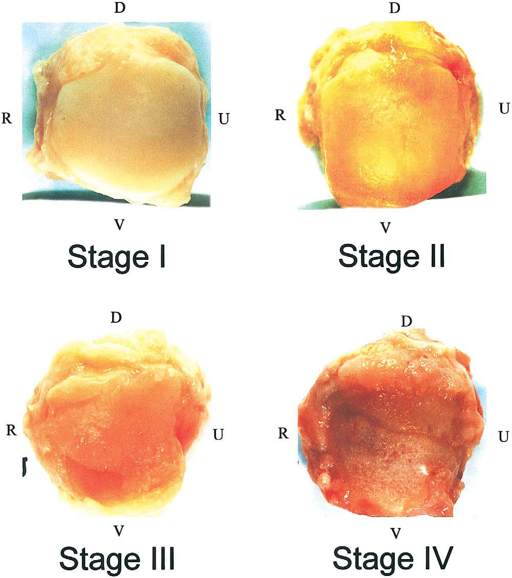
Human bite wounds contain very high concentrations of bacteria so the risk of infection is high. These infections can progress quickly and result in substantial complications, so early treatment is necessary (see Figure 2). Often, human bites occur when a person’s fist is driven into another’s mouth, such as during a fistfight. After the skin is broken, bacteria are seeded into the soft tissue and the ‘knuckle’ joint, which if left untreated often results in deep infection in the joint which may ultimately destroy the joint. These problems can be effectively treated by early diagnosis, intravenous antibiotics, and surgery to drain the infection out of the joint and wash it out.
Symptoms of Concern with Animal Bites to the HandIf the bite results in swelling, redness, warmth, continued pain beyond 24 hours, pus draining from the bite wound, red streaks extending up the arm or forearm, swollen lymph nodes (“glands”) around the elbow or in the armpit, loss of mobility, loss of sensation in the hand or fingertip, fever, malaise, night sweats, or rigors, emergency treatment should be sought either in your physician’s office or the emergency room.
Treatment of animal bitesYour doctor will examine the wound and ask about contributing factors to the injury. A complete history of the bite, including the type of animal and its status (general health, rabies vaccine, behavior), the time and location of the event, circumstances of the bite, whereabouts of the animal, and pre-hospital treatment will be reviewed.
It is crucial to update your tetanus status if you have not had a booster shot within the past ten years.
X-rays may be used to identify any damage to the bones and joints or tooth fragments that may have broken off. If an infected bite to the hand goes untreated for too long, x-rays may reveal evidence of osteomyelitis, or the spread of infection to the bone.
Animal bites to the hand require meticulous cleansing. Your doctor or other medical personnel will wash the wound and might trim away any devitalized (dead) tissue, damaged skin, blood clots, or other particles that could be a source of infection. It is important to look for signs of lymphangitis, indicated by the presence of red streaks on the forearm. Your doctor will feel the inner side of the elbow for evidence of enlarged lymph nodes. When the wound is infected, a culture is obtained to identify the type of bacteria that is causing the infection and thus help determine the antibiotic that is most effective for treatment.
The use of antibiotics for animal bites depends on the particular circumstances of the injury, patient health and sensitivity to various medications, and the appearance of the wound. Some bites require the use of IV antibiotics, while others may be treated with oral medication. The presence of an underlying fracture usually dictates inpatient antibiotic treatment. If you are diagnosed as having an infection of a flexor tendon sheath or a joint, you will need hand surgery, which will need to be performed as soon as possible.
Figure 1 Finger infection from cat bite

Figure 2. Wound Infection of thumb after human bite
Follow-up care is crucial in the case of animal bite wounds, to ensure that infection is diminishing or has not developed, and to restore the hand as much as possible to its former condition.

Carpal Boss

Clinical Characteristics


The dorsal wrist ganglion is most often confused with the carpal boss, so named by the French physician Foille.  The carpal boss is an osteoarthritic spur that develops at the base of the second and/or third carpometacarpal joints. (figure 1) A firm, bony, nonmobile, tender mass is visible and palpable at the base of the carpometacarpal joints, especially when the wrist is volar flexed.

Figure 1.


Radiologically, the mass is best visualized with the hand in 30 to 40 degrees supination and  

 20 to 30 degrees ulnar deviation ("carpal boss view")( figure 2).
Figure 2


The boss is more common in women (2:1), in the right hand (2:1), and between the third and fourth decades.  The mass may be asymptomatic, but the patient may complain of considerable pain and aching.  A small ganglion is associated with  the carpal boss in 30 percent of cases, adding to its confusion with the more common dorsal wrist ganglion. 

Injection to the ganglion or to the cmc joint may  be used to reduce pain and irritation.  This may be combined with splinting and anti inflammatory medication and avoidance of trauma to the back of the hand\
If symptoms persist at times surgery may be offered. (figure 2) Surgery may involve the removal of the prominent bone, the excision of an associated ganglion or cyst and at times involves tenosynovectomy or tenolysis of adjacent affected tendons.  

What occurs during surgery may depend upon the preoperative findings as well as the surgical intra operative findings
The most common complication is the persistence of a mass because of excision of the ganglion alone or inadequate excision of the osteophytes.  Pain will persist unless all abnormal abutting surfaces have been excised.  Dorsal wrist ganglions can present over the carpometacarpal joints and must be distinguished from the carpal boss with its own associated ganglion.  Avoidance of injury to branches of the radial and ulnar sensory nerves is again stressed.            

Arthritis of the Thumb and Wrist

What Is It?
In a normal joint, cartilage covers the end of the bones and serves as a shock absorber to allow smooth, pain-free movement. In osteoarthritis (OA, also called degenerative arthritis) the cartilage layer wears out, resulting in direct contact between the bones and producing pain and deformity. In the hand, one of the most common joints to develop OA is the base of the thumb. The thumb basal joint, also called the carpometacarpal (CMC) joint, is a specialized saddle-shaped joint that is formed by a small bone of the wrist (trapezium) and the first bone of the thumb (metacarpal).The saddle shaped joint allows the thumb its wide range of motions, including up, down, across the palm, and the ability to pinch (see Figure 1 below).
Figure 1.

Figure 2


Who Gets It?OA at the base of the thumb at the level of the wrist is more commonly seen in women over the age of 40.The exact cause is unknown, but genetics, previous injuries such as fractures or dislocations, and generalized joint laxity may predispose towards development of this type of arthritis.
What Are the Symptoms and Signs?The most common symptom is pain at the base of the thumb at the level of the wrist. The pain can be aggravated by activities that require pinch, such as opening jars, turning door knobs or keys, and writing. Also pain can progress to at rest and at night. In more severe cases, progressive destruction and mal-alignment of the joint occurs, and a bump develops at the base of the thumb, which occurs as the metacarpal moves out of the saddle joint. This shift in the joint can cause limited motion and weakness, making pinch difficult (see Figure 2).  The next joint above the CMC may compensate by loosening, causing it to bend further back (hyperextend).

How is the Diagnosis Made?

Figure 3
The diagnosis is made by history and physical evaluation. Pressure and movement such as twisting will produce pain at the joint.  A grinding sensation may also be present at the joint . Often a physician will perform a test called of all things, The Grind Test, to determine if loading the joint will cause pain.  Palpation or putting pressure on the joint  in selected areas is also used to diagnose this problem.  X-rays are used to confirm the diagnosis, although symptom severity often does not correlate with the x-ray findings. Often special X-ray views of the thumb and the wrist: including good images of the thumb and  trapezial views of the wrist are needed to fully delineate the arthritis.(see figure 3)


Figure 4. Base of the Thumb Metacarpal
sequential wear patterns arthritic stage (1)
The X-ray stage of the arthritis at the base of the thumb metacarpal has been shown to correlate with cartilage thickness and smoothness. ( see figure 4) Often in stage one it is very difficult to detect any xray changes despite changes to the articular cartilage. In stage II early joint spurs form at the edges of the joint.  These are typically less than 2mm in size. In stage III there is joint space narrowing as well as small joint spurs ans fragments.  Finally in stage IV arthritis  there is a narrow to often obliterated joint space and large bone spurs  know as osteophytes and changes to the subchondral bone ( bone just below the cartilage) often referred to as subchondral cysts and sclerosis or "whitening" of the bone on a standard xray image.


What are the Treatment Options?
Less severe thumb arthritis will usually respond to non-surgical care. Arthritis medication, splinting and  cortisone injections may help alleviate pain. A hand therapist might provide a variety of rigid and non-rigid splints which can be used while sleeping or during activities. However a rigid splint may irritate the skin if there are prominent bone spurs and can cause increase pain if too much immobilization occurs.
Recently topical medications such as a prescription anti-inflammatory  gel have had some success.
Patients with advanced disease or who fail non-surgical treatment may be candidates for surgical reconstruction.  A variety of surgical techniques are available that can successfully reduce or eliminate pain.  Surgical procedures vary and may include a combination of removal of arthritic bone and joint reconstruction , joint fusion, bone realignment, capsulodesis, tendon transfers, ligament reconstruction, release of contractures and even arthroscopy in very rare select cases. A consultation with your hand surgeon can help decide the best option for you.


Additional references

(1) Sequential Wear Patterns of the Articular Cartilage of the Thumb Carpometacarpal Joint in Osteoarthritis


Osteoarthritis of the Thumb Carpometacarpal Joint in Women and Occupational Risk Factors: A Case–Control Study

Diagnostic Value of Clinical Grind Test for Carpometacarpal Osteoarthritis of the Thumb


portions: © 2006 American Society for Surgery of the Hand and Modified/Addended by handctr for more see www.handctr.com

Tuesday, March 8, 2011

Facebook and THE HAND CENTER

The Hand Center of Western Massachusetts has updated its facebook site to include pages concerning Dupuytren's disease, arthritis, endoscopic carpal tunnel release and other interesting Hand related topics

Patient Choice Award 2010


Dr. Wint has been awarded a 2010 Patient Choice Award from www.vitals.com
This is the third year in a row that such an award has been given to him.

Monday, March 7, 2011

Golf Injuries to the Hand, Wrist or Elbow

The Hand Center of Western Massachusetts
Soon the snow will be gone and many in Western Massachusetts and Northern Connecticut will turn in their shovels and snowblowers for golf clubs. Golf can be especially hard on the hands, wrists and elbows. Make sure you are fit for this golf season.

Golf Injuries to the Hand, Wrist or Elbow

For most golfers, the hand and/or wrist is the third most common body region injured, after the back and elbow. The wrist is injured 3 times more frequently than the hand. In golf, the action of the wrist is important for the “snap” of the shot in long shots, and the precision “feel” in short shots.