Monday, November 21, 2011

OCD STICK snow blower safety this winter

Recommendations for safe use of a jammed snow blower snow blowers include : (OCD STICK) off, clutch, delay, stick

1. If the snow blower jams, immediately turn it OFF   
2. Disengage the CLUTCH
3.DELAY.. Wait 10 seconds after shutting of to allow Impeller Blades to stop rotating
4. Always use a STICK or broom handle to clear impacted snow. The stick most be strong enough to avoid breakage or eye injures can result from flying fragments
5. Never put your hand near chute or around blades
6. Keep all shields in place. Do not remove safety devices on machine
7. Keep hands and feet away from moving parts
8. Keep a clear head, concentrate and ...
        Do not drink alcoholic beverages before using a snow blower

As  physicians dedicated to the care of the Hand and Upper extremity we want to inform the public concerning the perils and pitfalls of improper snow blower use.  Physicians, nurses, allied health professionals and therapists who deal with these injuries live in fear of the first heavy wet snow of the season. Invariably injuries are seen despite general knowledge that these injuries occur. These safety tips cannot guarantee against injury but hopefully if you are reading these or even better spreading these... it is one more step towards preventing these types of injuries.

News organizations and weather services can help
Conditions that are associated with a higher incidence of injuries, hay wet snow exceeding 6 inches of accumulation and temperatures above 28 degrees Fahrenheit offer good opportunities to provide warning for the public. We need your help to reduce the incidence of these preventable injuries.


SNOWBLOWER SAFETY:  OFF CLUTCH DELAY STICK


ABOUT US: THE HAND CENTER

 The Hand Center

At The Hand Center of Western Massachusetts, we care for the hand, upper extremity, and shoulder, including fractures, tendonitis, lacerations, nerve compression, carpal tunnel, arthritis, and workers compensation injuries, just to name a few. 

The Hand Center is devoted to the care of the Hand, Upper Extremity and Shoulder. Our staff wants to provide you with a high quality of care. We are committed to successfully returning you to your daily work and leisure activities. 

We strive to work closely with our patients and their families, other medical professionals, employers and insurance carriers to provide you with needed care. We know that the path to recovery from minor to major injuries can be a difficult one. Our highly specialized physicians and caring staff are interested in your success. 
Our office is unique in Western New England where orthopedic surgeons and plastic surgeons often work in the field of Hand Surgery but ours is the only practice in the Pioneer Valley that is devoted exclusively to the Hand and Upper Extremity.  In New England Orthopedic Surgeons and Plastic Surgeons are often in busy offices that may not have the attention to fine details of your Hand and Upper Extremity needs.  At the Hand  Center of  Western Massachusetts we pride ourselves in this facet of your care.  So whether you are having a new problem or have been all over the area and have seen  other New England area Orthopedic or Plastic Surgeons seeking care for an upper extremity problem we are happy to consider seeing you in consultation.  

Here at The Hand Center of Western Massachusetts our physicians provide the individual attention and care that you need. You will always be seen by one of our highly skilled Hand surgeons at every visit.  Our staff and physicians often treat complex problems seen by other Orthopedic Surgeons in the New England area, and we feel that if you are coming to see one of us you should see a physician each and every time.

Doctors Wint, Wintman, and Martin are fellowship trained hand surgeons. Doctors Wint and Wintman are trained in and Board Certified in Orthopaedic surgery with certificates of added qualifications in surgery of the hand.  Dr. Martin is Board certified trained in plastic surgery, and has a certificate in hand surgery as well

Our physicians have surgical privileges at Baystate Medical Center, Pioneer Valley SurgiCenter, Mercy Medical Center, Mary Lane Hospital,  Cooley Dickinson Hospital and Noble Hospital

WHAT to do after a hand injury and an ER visit?

WHAT SHOULD I DO IF I HAVE BEEN SEEN AT A HOSPITAL FOR A FINGER, HAND, WRIST or FOREARM INJURY?


Q. What should I do If I've been seen in a local emergency room for a hand, wrist, forearm or upper extremity fracture or injury?
A. It is important that day or night to follow the ER or ED instructions for post injury care. If you are told to elevate or ice or take a certain medication please do as you are instructed.  Make sure you fill prescriptions  that you may need.  If you are not sure whether a medicine given to you by the Emergency Physician is correct call the hospital back and ask.  In addition you should arrange for appropriate follow up care with a qualified physician.
Here are 6 important TIPS for delayed and urgent care of HAND AND WRIST FRACTURES that have been seen in a local Emergency Department ...in Western Massachusetts and Northern Connecticut
1.Contact your PCP. They can provide interim care or redirect you.
2. Call us. We like to accommodate potential patients. Unfortunately we cant always accommodate everyone who calls.
3. Call the ER back. Often they can advise you over the phone if you've been seen recently
4. Return to the ER where you were seen. Most ER's will be glad to see you again
5. Call  a doctor you know of on your own. There is nothing wrong with seeking care on your own
6. Don't panic. Many hand and wrist injuries once stabilized in a local ER can be seen safely and effectively at a later date.
Most Hospital Emergency Departments will have given you a follow-up for a physician that they regularly work with, THERE ARE SOME TIMES THAT THEY DON'T HAVE A HAND SURGEON ON CALL OR ON STAFF. IN THOSE CASES YOU SHOULD SEEK OUT A HAND SURGERY GROUP IN THE AREA. WHILE THERE MAY NOT BE AN IMMEDIATE APPOINTMENT AVAILABLE RECORDS FROM YOUR ER VISIT CAN BE OBTAINED AND IN MANY CASES AN APPOINTMENT CAN BE GIVEN WITHIN THE PROPER TIME FRAME. IT IS IMPORTANT TO REMEMBER THAT WHILE THE ER MAY STRESS BEING SEEN IMMEDIATELY THAT MANY HAND AND WRIST CONDITIONS CAN BE TAKEN CARE OF URGENTLY BUT NOT EMERGENTLY ONCE PROPER ER EMERGENT CARE HAS BEEN GIVEN. However there are conditions that may warrant being seen right away and at times you may be advised to go back to the original ER if you cant be seen in an adequate time frame. Every situation is different. and this information is not to be construed as ultimate medical advice. for the purpose of treatment. 
 

Fireworks Safety

Hand Surgeons Agree:

Leave Fireworks to the Professionals 


The American Society for Surgery of the Hand (ASSH) has urged the public to leave fireworks in the hands of the professionals.According to the U.S. Consumer Product Safety Commission, 38% of all reported fireworks-related injuries from June 22-July 22, 2001, were to fingers, hands, and arms. These injuries included burns, lacerations, fractures, and traumatic amputation.
Of the finger, hand, and arm injuries, the majority of injuries were caused from accidents involving firecrackers, bottle rockets, and sparklers— the three firework-types most often used in a backyard environment. Accidents involving firecrackers, bottle rockets, and hand-held sparklers totaled 57% of all firework injuries (source: American Pyrotechnic Association).
One solution that has been offered by the ASSH to individuals is to attend public fireworks displays, which are monitored for safety by a local fire department, rather than setting off fireworks near or around the home.
The following precautions should be taken when attending a public fireworks display:
  • Obey safety barriers and ushers.
  • Stay back a minimum of 500 feet from the launching site.
  • Resist the temptation to pick up firework debris when the display is over. The debris may still be hot, or in some cases, the debris might be “live” and could still explode.
  • Never give children hand-held sparklers. Sparklers cause 10% of all firework injuries (source: American Pyrotechnics Association)—and were associated with the most injuries to children under 5 years of age. (source: U.S. Consumer Product Safety Commission)


                                                        Keep your hands safe this fourth of July. Enjoy the day and leave fireworks to the professionals
 
                                                                                                               www.nfpa.org
                     Their PSA features voices of victims whose lives have been tragically altered due to fireworks

portions Copyright © American Society for Surgery of the Hand 2008.
Modified/adapted altered by www.handctr.com from www.assh.org

GOUT (from medscape)

GOUT (from medscape)

  • Gout has 2 clinical phases: (1) a first phase of intermittent acute attacks that spontaneously resolve during 7 to 10 days with asymptomatic periods between attacks, and (2) a second phase of chronic tophaceous gout involving polyarticular attacks with crystal deposition (tophi) in the soft tissues or joints.
  • Risk factors include use of thiazide diuretics, cyclosporine, and low-dose aspirin (< 1 g/day); insulin resistance metabolic syndrome; renal insufficiency; hypertension; congestive heart failure; and organ transplantation.
  • Increased dietary intake of purines, ethanol, soft drinks, and fructose also increase the risk for gout. Intake of coffee, dairy products, and vitamin C reduces the risk for gout.
  • Triggers for gout attacks include alcohol intake, diuretic use, hospitalization, and surgery.
  • The diagnostic standard is synovial fluid examination for negatively birefringent monosodium urate crystals under polarizing microscopy.
  • Hyperuricemia may not be present in an acute attack and may not be helpful in diagnosis.
  • The differential diagnosis of acute gout includes other crystal-induced arthritides, rheumatoid arthritis, and a septic joint.
  • The main aim of treatment is rapid pain relief and prevention of disability.
  • Options include the use of NSAIDs; colchicine; glucocorticoids; and, sometimes, corticotrophin.
  • Adjunctive measures include applying ice and resting the affected joint.
  • NSAIDs and colchicine are first-line treatments of acute gout.
  • Colchicine, given at 1.2 mg at the start of an attack and repeated at 0.6 mg 1 hour later, is more effective than placebo for pain relief within 24 hours.
  • Glucocorticoids and corticotrophins may be used, but the evidence for intramuscular injections is limited.
  • A 5-day course of prednisolone has been shown to be equivalent to indomethacin and naproxen.
  • 7 to 10 days of treatment of gout may be needed for symptom control.
  • Lowering urate levels may prevent acute flares of gout and development of tophi.
  • Urate-lowering therapy for hyperuricemia is recommended for those with at least 2 gout attacks per year or tophi, but such therapy should not be initiated during acute attacks.
  • Urate-lowering therapy should be started 2 to 4 weeks after flare resolution, with a low initial dose increased for weeks to months.
  • The dose should be adjusted to achieve a urate level below 6 mg/dL, which is associated with a reduced risk for acute attacks and tophi.
  • Allopurinol, a xanthine oxidase inhibitor, is the most commonly prescribed agent to lower urate levels.
  • Febuxostat is another xanthine oxidase inhibitor approved by the FDA in 2009. At daily doses of 80 mg and 120 mg, has been shown to be 2.5 to 3 times more likely to achieve urate levels less than 6 mg/dL at 1 year.
  • Uricosuric drugs (probenecid, sulfinpyrazone, and benzbromarone) block renal tubular urate reabsorption.
  • Uricase and pegloticase were FDA approved in 2010 for chronic gout refractory to conventional treatments.
  • Lifestyle changes such as avoidance of alcohol and diet modification may not be sufficient to control attacks.
  • Intake of vitamin C and dairy products have been shown to reduce urate levels.
  • For patients starting allopurinol therapy, the use of colchicine at a dose of 0.6 mg twice daily for an average of 5.2 months has been shown to reduce the risk for gout attacks and severity of flares.
  • The optimal duration of treatment of tophi is uncertain, and ongoing prophylaxis until resolution of tophi occurs may be necessary.
  • The author concluded that patients suspected of having gout should have the diagnosis confirmed by synovial fluid examination, and acute treatment after multiple attacks should be followed by allopurinol for urate lowering, with colchicine prophylaxis to prevent recurrence of attacks.


WORK RELATED CARPAL TUNNEL IN MASSACHUSETTS

WORK RELATED CARPAL TUNNEL IN MASSACHUSETTS FACT SHEET

What is Carpal Tunnel Syndrome (CTS)?
Carpal Tunnel Syndrome (CTS) involves compression of the median nerve at the wrist. The finger tendons, blood vessels and median nerve extend from the forearm to the fingers through a small tunnel (surrounded by bone) in the wrist, named the carpal tunnel. If any of the tendons in the carpal tunnel become swollen, the median nerve is pinched resulting in pain, numbness and tingling of the first three fingers of the hand. If CTS is not treated in its early stages, it can result in permanent disability. It is estimated that close to 1 million people in the United States annually may develop CTS, requiring medical care and leaving them at least temporarily disabled.1

What Causes CTS?
CTS can be caused by chronic diseases such as rheumatoid arthritis, gout, and diabetes. It has also been linked with pregnancy and birth control use. Exposure to workplace factors can also cause CTS. It is estimated that approximately 50% of all medically diagnosed cases of CTS are work-related.2,3

Work-related risk factors for CTS include repetitive and forceful exertions of the hands and wrists, combinations of either force or repetitive work and awkward hand postures, and exposure to hand vibration.4 Acute trauma to the wrist can also cause CTS.

Is Work-Related CTS Preventable?
Work related CTS (WR-CTS) is preventable. Prevention practices to reduce the risk of developing CTS are varied. Examples are:

•Adjusting your workstation so that you are working in the proper posture;
•Reducing the number of times per hour or a day you use your fingers and hands;
•Reducing the number and weight of forceful exertions such as lifting or pinching objects.
•Using well maintained, correctly sized tools.
Other prevention strategies involve changing work organization. These include, for example, reducing the pace of work, and making certain you take frequent rest breaks throughout the day.

What is known about work-related CTS in Massachusetts?
Since 1992, the Massachusetts Department of Public Health (MDPH), funded by the National Institute of Occupational Safety and Health, has conducted surveillance of work-related CTS in order to identify industries, occupations and workplaces where prevention efforts are needed. The Occupational Health Surveillance Program (OHSP) at the Department uses two data sources to identify cases of work-related CTS: 1) workers' compensation claims filed in Massachusetts with an injury code for "CTS"; and 2) case reports of confirmed and suspected work-related CTS filed by physicians in accordance with Massachusetts regulations requiring physicians to report select work-related conditions to MDPH.

Key Surveillance Findings
•Between March 1992 and June 1997, 4,837 cases of work-related-CTS were reported to OSHP.
•The highest rates of work-related CTS were found among workers in manufacturing industries. However, the largest numbers of cases were employed in technical, sales and administrative support occupations. Fifteen percent of the cases employed in manufacturing were employed in administrative/sales jobs.
•Data entry keyers, general office clerks, secretaries and cashiers appear on the list of occupations with both high numbers of cases and high rates of work-related CTS.
•Grocery stores were the single industry with the highest number of cases; cashiers made up 40% of the cases. Hospitals (a service industry) has the second highest number of cases. Secretaries make up 15% of the cases in the hospital industry.
•Over 300 cases were less than 25 years old, raising significant concern about the long term impact of work-related CTS on health and employment options of young workers who are just beginning their careers.
•Many more women in Massachusetts are getting work-related CTS than men. A number of factors likely account for this finding. Women may be more likely to select or be selected into high risk jobs. Underlying biological differences between men and women and gender differences in reporting injuries and seeking medical care are also possible factors.
•Over 70% of cases identified through workers' compensation who were interviewed have had surgery for their CTS and almost 70% reported CTS in both hands.
•Approximately 50% of the workers who attributed their CTS to keyboard use reported having had surgery for CTS and 50% reported CTS in both hands

1 Tanaka S, Wild D, Seligaman P, Behrens V, Cameron L and Putz-Anderson V.(1994):The US prevalence of self-reported carpal tunnel syndrome: 1988 national health interview survey data. Am J of Public Health 84(11):1846-1848.

2 Ibid.

3 Cummings H, Maizlish N, Rudolph L, Dervin K, Ervin A (1989): Occupational disease surveillance: Carpal tunnel syndrome. Morbidity and Mortality Weekly Report 38: 485-489.

4 Bernard B (1997): "Musculoskeletal disorders and workplace factors: A critical review of epidemiologic evidence for work-related musculoskeletal disorders of the neck, upper extremity, and low back." Cincinnati: Department of Health and Human Services, National Institute of Occupational Safety and Health

SOURCE

MALLET FINGER____BASEBALL FINGER


MALLET FINGER (BASEBALL FINGER)


A mallet finger occurs when the extensor tendon at the tip of a finger ruptures. The rupture of this tendon can involve the tendon alone, be associated with a small bone fragment or fracture or can be associated with a fracture that requires significant care.

The force applied to the finger can come from something as simple as tucking in a bed sheet or can come from a direct blow to the end of a finger. Mallet finger has also been known as baseball finger.

A mallet finger often begins with pain at the distal joint of the finger.  At times there is an immediate loss of motion while at other times the finger seems to stay straight for a while and only later starts to lose its ability to be extended actively at the tip.  At times there is an injuries are typically closed in that the skin and nail is intact but at times there is an injury to the skin or nail bed as well. In severe cases the injury is associated with an open injury to the joint or bone, a so called open or compound fracture.

In adults the injury can involve the joint surface.  In children it can involves the growth plate or physis.

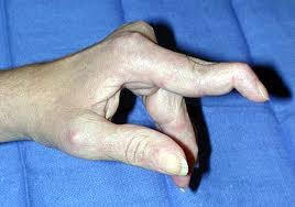
The diagnosis is often made based upon the type of injury and the appearance of the finger.  The fingertip will droop down and there is a loss of active motion.  Often the finger can be passively pushed up to straighten it but the independent active motion to extend the digit at the tip has been lost

Mallet finger.
X-rays are often taken to further delineate the injury and see how much if any bone, joint or growth plate is involved

Treatment depends largely upon the extent the soft tissue and underlying boney injury.

Tendon rupture without bone injury

Most of these mallet finger injuries can be treated with splinting.  The splint can be applied in a variety of ways depending upon the injury.  Typically the split is left in place full time for six to eight weeks with a time for part time splinting after that depending upon what daily activity is done by the patient with a typical part time period of 3 – 4 weeks.  In some situations pinning of the joint is used rather than a splint

Tendon rupture with a small bone fragment

These injuries typically are treated like non-boney injuries
Types of splints used to treat mallet finger. A, Dorsal aluminum splint. B, Commercial splint.
Reproduced with permission from Culver JE Jr: Office management of athletic injuries of the hand and wrist. Instr Course Lect 1989;38:473-482.

Tendon rupture with a large bone fragment involving the joint.


These injuries may respond to splinting and splinting is often used however a small bump may always be present a t the joint.  At times if the doctor feels that that the bone fragment is large enough and the joint may be unstable surgery may be offered.  During surgery pins or small screws may be used and the joint itself may be pinned to prevent motion during the healing process.

Above: X-rays showing fracture at the insertion of the extensor tendon. In the first image on the left the fragment is displaced.  This will heal with a bump but will be able to be treated with a splint.  IN the image on the right the joint has subluxed.  This will  need to have surgical repair.
In adults with severe open injury more immediate surgery may also be offered

Children

In children the doctor needs to differentiate between these injuries that require reduction or realignment of the bone without surgery and those who may have a portion of the nail bed significantly torn or retained within the fracture site or growth plate.  Often children will not have a tendon injury but a fracture through the physis which appears to be a mallet injury.  X-rays often will reveal this.


Mallet deformity from a fracture across the growth plate in a child is different than the adult fracture or tendon avulsion

Late or Delayed Treatment in adults

Delayed treatment of mallet finger deformity may consist of splinting initially and at times surgical methods are offered to correct chronic deformities and other associated joint and tendon problems that may accompany the chronic situation

Results

Most mallet fingers heal well, although often there is a slight loss of full extension. The slight extension loss typically has no effect on hand of finger function, but if left untreated it can cause other issues to occur in the finger due to tendon imbalance. While treatment of a closed mallet finger is not an acute emergency, the improper, partial or untreated injury can lead to further problems such as a swan neck deformity.

 Swan neck deformity from tendon imbalance and laxity at the proximal joint.
 
Athletics
While there may be special circumstances where a professional athletes plays with a splint in place for mallet finger for the child or recreational athlete, or even most professional or collegiate level athletes this is not typically recommended.

CARPAL BOSS of the HAND and WRiST

Carpal Boss
  
Clinical Characteristics
The dorsal wrist ganglion is most often confused with the carpal boss, so named by the French physician Foille.  The carpal boss is an osteoarthritic spur that develops at the base of the second and/or third carpometacarpal joints. (figure 1) A firm, bony, nonmobile, tender mass is visible and palpable at the base of the carpometacarpal joints, especially 

when the wrist is volar flexed. The lining of the joint thins out and small bone spurs form n the top of the hand leadingto a bone prominence. Tendon irritation can occur and a small cyst or ganglion may form as well.


figure 1 Right hand with carpal boss


Figure 2 Carpal Boss seen on Xray
Radiologically, the mass is best visualized with the hand in 30 to 40 degrees supination and  
 20 to 30 degrees ulnar deviation ("carpal boss view")( figure 2).
The boss is more common in women (2:1), in the right hand (2:1), and between the third and fourth decades.  The mass may be asymptomatic, but the patient may complain of considerable pain and aching.  A small ganglion is associated with  the carpal boss in 30 percent of cases, adding to its confusion with the more common dorsal wrist ganglion. 


Injection to the ganglion or to the cmc joint may  be used to reduce pain and irritation.  This may be combined with splinting and anti inflammatory medication and avoidance of trauma to the back of the hand\

If symptoms persist at times surgery may be offered. (figure 3) Surgery may involve the removal of the prominent bone, the excision of an associated ganglion or cyst and at times involves tenosynovectomy or tenolysis of adjacent affected tendons.  What occurs during surgery may depend upon the preoperative findings as well as the surgical intra operative findings
Figure 3  Surgical Approach to carpal boss excision using a transverse incision
The most common complication is the persistence of a mass because of excision of the ganglion alone or inadequate excision of the osteophytes.  Pain will persist unless all abnormal abutting surfaces have been excised.  Dorsal wrist ganglions can present over the carpometacarpal joints and must be distinguished from the carpal boss with its own associated ganglion.  Avoidance of injury to branches of the radial and ulnar sensory nerves is again stressed.            
Endoscopic Carpal Tunnel Release Surgery
visit the Facebook page

   
Endoscopic Carpal Tunnel Release Surgery

fireworks safety stressed

Fireworks danger stressed

Thursday, July 01, 2004
By NANCY H. GONTER
ngonter@repub.com

NORTHAMPTON - Hand surgeon Jeffrey Wint isn't expecting a quiet Fourth of July weekend.
 
 
"Unfortunately, I face the prospect of being called in at any time," said Wint, who hopes to take in a parade and spend some time in his back yard during the holiday.
Bookmark and Share
Wint, other medical professionals, law enforcement and fire officials yesterday held a press conference calling on area residents to "leave fireworks to the professionals." Members of the state police bomb squad exploded illegal fireworks using mannequins to show the damage they can cause.

A surgeon at the Hand Center of Western Massachusetts who works at Baystate Medical Center in Springfield, Wint said he has already seen hand injuries caused by fireworks this season.

In one case, a teenage boy was clenching a firecracker in his hand and it blew up before he threw it. The tip of his index finger had to be amputated. In another case, a 14-year-old boy didn't realize the cherry bomb in his hand was lit. He suffered burns to his palm, deep lacerations and several fractures.

"He has the prospect of months of therapy and rehabilitation in order to regain normal function," Wint said.

Of injuries to the hand and upper extremities, 57 percent are caused by fireworks, Wint said.

Patrick C. Lee, a trauma surgeon at Baystate Medical Center, said burn injuries are tragic for children and their families.

"They are a life-changing event. Burns and their treatment are painful, leave surgical and emotional scars, and most of all they are preventable," Lee said.

State Fire Marshal Stephen D. Coan said many people ask him why children shouldn't play with sparklers.

"I ask them if they would give their children a lighter to play with. Sparklers can burn at 1,800 degrees Fahrenheit, three times the temperature of a lit match, and hot sparkler fires have ignited clothing, burned bare feet, poked eyes out and started tragic holiday fires," Coan said.

Coan noted that all fireworks are illegal in Massachusetts and it is illegal to bring fireworks into the state, even though they are legal in Connecticut and New Hampshire. It is also illegal to purchase fireworks on the Internet or through catalogs and have them delivered to homes in Massachusetts, he said.

"Remember, the use of fireworks by anyone other than a licensed professional is illegal," Coan said.

The press conference featured a table loaded with fireworks in colorful packaging, much of it clearly intended to attract children. The fireworks were seized by law enforcement authorities. Coan noted children 10-14 are at the greatest risk of fireworks injuries.

Fireworks can be deadly. In December 2003, a 45-year-old Gloucester woman died when fireworks ignited her Christmas tree, starting a house fire. In May 1997, a 26-year-old Watertown man was killed when lighting fireworks in a hallway. On July 4, 1993, a 27-year-old Framingham man was killed when his backyard fireworks exploded in his face, according to the state Department of Fire Services.

MORE ON FIREWORKS

Fireworks safety


Fireworks Safety Brief
LOSSES
According to the Consumer Product Safety Commission, national losses involving fireworks amount to 3 deaths and 10,527 injuries annually. Hand and finger injuries are the most common and account for 32 percent of all injuries. Head and eye injuries occur with about the same frequency, equaling 19 and 18 percent of total injuries.
BACKGROUND
A review of firework mishaps shows a variety of factors contribute to the typical mishap. Most pre-school age victims are injured by fireworks ignited by someone else, while older children who are injured are usually lighting the fireworks themselves. Children under age five are commonly hurt by rocket-type fireworks; small firecrackers and ground spinners injure the majority of children between the ages of 5 and 14. Most of the injuries associated with large, illegal firecrackers such as M-80's are to older teenagers or adults.
Bookmark and Share
 
FOR MORE INFORMATION concerning fireworks safety go to these links below.

NFPA--FireworksSafetyTips.pdf
AAOS press release
ASSH fireworks 2006 press release
Press Release Summer 2004
Channel 22 WWLP Springfield , 6/30/04 
Springfield Republican Article 7/1/04
http://www.fireworksafety.com/statelaws/index.html


Click to visit our website

Flexor Tendon Injuries 

Flexor tendons in the hand and forearm

The muscles that bend (flex) the fingers are called flexor muscles. These flexor muscles move the fingers through cord-like extensions called tendons, which connect the muscles to bone. The flexor muscles start at the elbow and forearm regions, turn into tendons just past the middle of the forearm, and attach to the bones of the fingers (see Figure 1). In the finger, the tendons pass through fibrous rings called pulleys, which guide the tendons and keep them close to the bones, enabling the tendons to move the joints much more effectively.

Deep cuts on the palm side of the wrist, hand, or fingers can injure the flexor tendons and nearby nerves and blood vessels. The injury may appear simple on the outside, but is actually much more complex on the inside. When a tendon is cut, it acts like a rubber band, and its cut ends pull away from each other. A tendon that has not been cut completely through may still allow the fingers to bend, but can cause pain or catching, and may eventually tear all the way through. When tendons are cut completely through, the finger joints cannot bend on their own (see Figure 2).The Hand that has a ruptured or cut flexor tendon will not have a normal pattern or cascade to the finger tips. Observation of the hand will reveal that the finger with a cut or ruptured flexor tendon will "stick out" due to the absence of normal resting tension (see Figure 3)
Bookmark and Share

How are flexor tendon injuries treated?

Tendon Healing

Tendons are made of living cells. If the cut ends of the tendon can be brought back together, healing begins through the cells that are inside of the tendon as well as the tissue outside of the tendon. Because the cut ends of a tendon usually separate after an injury, a cut tendon can not heal without surgery.

Your doctor will advise you on how soon surgery is needed after a flexor tendon is cut. There are many ways to repair a cut tendon, and certain types of cuts need a specific type of repair. In the finger, it is important to preserve certain pulleys, and there is very little space between the tendon and pulley in which to perform a repair. Nearby nerves and blood vessels may need to be repaired as well. After surgery, and depending on the type of cut, the injured area can either be protected from movement or started on a very specific limited-movement program for several weeks (see Figure 4). Your doctor may prescribe hand therapy for you after surgery. If unprotected finger motion begins too soon, the tendon repair is likely to pull apart. After four-to-six weeks, the fingers are allowed to move slowly and without resistance. Healing takes place during the first three months after the repair.

In most cases, full and normal movement of the injured area does not return after surgery. If it is hard to bend the finger using its own muscle power, it could mean that the repaired tendon has pulled apart or is bogged down in scar tissue. Scarring of the tendon repair is a normal part of the healing process. But in some cases, the scarring can make bending and straightening of the finger very difficult. Depending on the injury, your doctor may prescribe therapy to loosen up the scar tissue and prevent it from interfering with the finger’s movement. If therapy fails to improve motion, surgery to release scar tissue around the tendon may be required.

Hand Therapy After Surgery
If a program of controlled, limited motion is selected as therapy for the first several weeks after surgery, it is important to work closely with a hand therapist and your surgeon to understand the therapy and follow set guidelines. The tendon repair might pull apart if your hand is used too soon or if therapy guidelines are not followed. In addition to regaining motion of the finger after a tendon injury, therapy will be helpful in softening scars and building grip strength.


Figure 1: The tendons of the hand run from the flexor muscles in the elbow and forearm to the bones of the fingers.

Figure 2: When flexor tendons are completely cut, the finger cannot be bent.


Figure 3: After a flexor tendon rupture or laceration the affected finger will often stick out and not follow the normal cascade or posture of the resting hand.

Figure 4: After surgery, the area of the injured tendon must be protected from movement.

Portions © 2009 American Society for Surgery of the Hand. Developed by the ASSH Public Education Committee
Taken, modiifed and adapted by www.handctr.com from www.assh.org ( ASSH)
Portions © from www.handctr.com
flexor_injuries.pdf